Traduzione e analisi delle parole tramite l'intelligenza artificiale ChatGPT
In questa pagina puoi ottenere un'analisi dettagliata di una parola o frase, prodotta utilizzando la migliore tecnologia di intelligenza artificiale fino ad oggi:
- come viene usata la parola
- frequenza di utilizzo
- è usato più spesso nel discorso orale o scritto
- opzioni di traduzione delle parole
- esempi di utilizzo (varie frasi con traduzione)
- etimologia
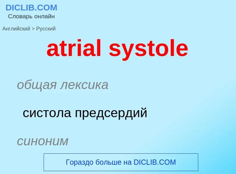
atrial systole - traduzione in Inglese


![CGI animated]] graphic of the human heart, sectioned, with motions and timing synced with the Wiggers diagram. The section shows: 1) the opened ventricles contracting once per heartbeat—that is, once per each cardiac cycle; 2) the (partly obscured) mitral valve of the left heart; 3) the tricuspid and pulmonary valves of the right heart—note these paired valves open and close oppositely. + (The aortic valve of the left heart is located below the pulmonary valve, and is completely obscured.) The (unsectioned) atria are seen above the ventricles.](https://commons.wikimedia.org/wiki/Special:FilePath/Cardiac-Cycle-Animated.gif?width=200 "CGI animated]] graphic of the human heart, sectioned, with motions and timing synced with the Wiggers diagram. The section shows: 1) the opened ventricles contracting once per heartbeat—that is, once per each cardiac cycle; 2) the (partly obscured) mitral valve of the left heart; 3) the tricuspid and pulmonary valves of the right heart—note these paired valves open and close oppositely. + (The aortic valve of the left heart is located below the pulmonary valve, and is completely obscured.) The (unsectioned) atria are seen above the ventricles.")
, ''mitral'' in the left heart (pink)) are open to enable blood to flow directly into both left and right ventricles, where it is collected for the next contraction.")
![Cardiac (ventricular) systole: Both AV valves (''tricuspid'' in the right heart (light-blue), ''mitral'' in the left heart (pink)) are closed by back-pressure as the ventricles are contracted and their blood volumes are ejected through the newly-opened ''pulmonary valve'' (dark-blue arrow) and ''aortic valve'' (dark-red arrow) into the [[pulmonary trunk]] and aorta respectively.](https://commons.wikimedia.org/wiki/Special:FilePath/Heart systole.svg?width=200 "Cardiac (ventricular) systole: Both AV valves (''tricuspid'' in the right heart (light-blue), ''mitral'' in the left heart (pink)) are closed by back-pressure as the ventricles are contracted and their blood volumes are ejected through the newly-opened ''pulmonary valve'' (dark-blue arrow) and ''aortic valve'' (dark-red arrow) into the [[pulmonary trunk]] and aorta respectively.")
![The parts of a [[QRS complex]] and adjacent deflections. Re the cardiac cycle, ''atrial systole'' begins at the P wave; ''ventricular systole'' begins at the Q deflection of the QRS complex.](https://commons.wikimedia.org/wiki/Special:FilePath/QRS normal.svg?width=200 "The parts of a [[QRS complex]] and adjacent deflections. Re the cardiac cycle, ''atrial systole'' begins at the P wave; ''ventricular systole'' begins at the Q deflection of the QRS complex.")
 normally refers to atria and ventricles at relaxation and expansion together—while refilling with blood returning to the heart. Systole (left) typically refers to ''ventricular systole'', during which the ventricles are pumping (or ejecting) blood out of the heart through the aorta and the pulmonary veins.")
![A [[Wiggers diagram]] illustrate events and details of the cardiac cycle with electrographic trace lines, which depict (vertical) changes in a parameter's value as time elapses left-to-right. The ventricular "Diastole", or relaxation, begins with "Isovolumic relaxation", then proceeds through three sub-stages of inflow, namely: "Rapid inflow", "Diastasis", and "Atrial systole". (During the "Diastole" period, the "Ventricular volume" increases (see red-line tracing), beginning after the vertical bar at ''"Aortic valve closes"'' and ending with the vertical bar at R in the QRS complex). + The ventricular "Systole", or contraction, begins with "Isovolumic contraction", i.e., with the vertical bar at ''"A -V valve closes"''; it ends with completing the "Ejection" stage at the bar at ''"Aortic valve closes"''. During "Ejection" stage, the (red-line) tracing of "Ventricular volume" falls to its least amount (see [[ejection fraction]]) as the ventricles pump blood to the pulmonary arteries and to the aorta.](https://commons.wikimedia.org/wiki/Special:FilePath/Wiggers Diagram 2.svg?width=200 "A [[Wiggers diagram]] illustrate events and details of the cardiac cycle with electrographic trace lines, which depict (vertical) changes in a parameter's value as time elapses left-to-right. The ventricular \"Diastole\", or relaxation, begins with \"Isovolumic relaxation\", then proceeds through three sub-stages of inflow, namely: \"Rapid inflow\", \"Diastasis\", and \"Atrial systole\". (During the \"Diastole\" period, the \"Ventricular volume\" increases (see red-line tracing), beginning after the vertical bar at ''\"Aortic valve closes\"'' and ending with the vertical bar at R in the QRS complex). + The ventricular \"Systole\", or contraction, begins with \"Isovolumic contraction\", i.e., with the vertical bar at ''\"A -V valve closes\"''; it ends with completing the \"Ejection\" stage at the bar at ''\"Aortic valve closes\"''. During \"Ejection\" stage, the (red-line) tracing of \"Ventricular volume\" falls to its least amount (see [[ejection fraction]]) as the ventricles pump blood to the pulmonary arteries and to the aorta.")
общая лексика
систола предсердий
синоним
общая лексика
систола желудочков
общая лексика
сердечный цикл
Definizione
Wikipedia

The cardiac cycle is the performance of the human heart from the beginning of one heartbeat to the beginning of the next. It consists of two periods: one during which the heart muscle relaxes and refills with blood, called diastole, following a period of robust contraction and pumping of blood, called systole. After emptying, the heart relaxes and expands to receive another influx of blood returning from the lungs and other systems of the body, before again contracting to pump blood to the lungs and those systems. A normally performing heart must be fully expanded before it can efficiently pump again. Assuming a healthy heart and a typical rate of 70 to 75 beats per minute, each cardiac cycle, or heartbeat, takes about 0.8 second to complete the cycle. There are two atrial and two ventricle chambers of the heart; they are paired as the left heart and the right heart—that is, the left atrium with the left ventricle, the right atrium with the right ventricle—and they work in concert to repeat the cardiac cycle continuously (see cycle diagram at right margin). At the start of the cycle, during ventricular diastole–early, the heart relaxes and expands while receiving blood into both ventricles through both atria; then, near the end of ventricular diastole–late, the two atria begin to contract (atrial systole), and each atrium pumps blood into the ventricle below it. During ventricular systole the ventricles are contracting and vigorously pulsing (or ejecting) two separated blood supplies from the heart—one to the lungs and one to all other body organs and systems—while the two atria are relaxed (atrial diastole). This precise coordination ensures that blood is efficiently collected and circulated throughout the body.
The mitral and tricuspid valves, also known as the atrioventricular, or AV valves, open during ventricular diastole to permit filling. Late in the filling period the atria begin to contract (atrial systole) forcing a final crop of blood into the ventricles under pressure—see cycle diagram. Then, prompted by electrical signals from the sinoatrial node, the ventricles start contracting (ventricular systole), and as back-pressure against them increases the AV valves are forced to close, which stops the blood volumes in the ventricles from flowing in or out; this is known as the isovolumic contraction stage.
Due to the contractions of the systole, pressures in the ventricles rise quickly, exceeding the pressures in the trunks of the aorta and the pulmonary arteries and causing the requisite valves (the aortic and pulmonary valves) to open—which results in separated blood volumes being ejected from the two ventricles. This is the ejection stage of the cardiac cycle; it is depicted (see circular diagram) as the ventricular systole–first phase followed by the ventricular systole–second phase. After ventricular pressures fall below their peak(s) and below those in the trunks of the aorta and pulmonary arteries, the aortic and pulmonary valves close again—see, at the right margin, Wiggers diagram, blue-line tracing.
Now follows the isovolumic relaxation, during which pressure within the ventricles begin to fall significantly, and thereafter the atria begin refilling as blood returns to flow into the right atrium (from the vena cavae) and into the left atrium (from the pulmonary veins). As the ventricles begin to relax, the mitral and tricuspid valves open again, and the completed cycle returns to ventricular diastole and a new "Start" of the cardiac cycle.
Throughout the cardiac cycle, blood pressure increases and decreases. The movements of cardiac muscle are coordinated by a series of electrical impulses produced by specialised pacemaker cells found within the sinoatrial node and the atrioventricular node. Cardiac muscle is composed of myocytes which initiate their internal contractions without applying to external nerves—with the exception of changes in the heart rate due to metabolic demand.
In an electrocardiogram, electrical systole initiates the atrial systole at the P wave deflection of a steady signal; and it starts contractions (systole).